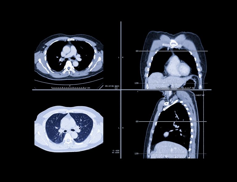
Last year, there were 1.8 million deaths from lung cancer worldwide according to estimates from the World Health Organization. Despite a decrease in the number of people smoking and advances in early detection and treatment new cases of lung cancer continue to rise and is the leading cause of cancer death among men and women. In fact, each year more people die from lung cancer than from colon, breast and prostate cancers combined.
But a new advance in early lung cancer detection that involves artificial intelligence (AI), provides another tool in the arsenal of the fight against this deadly disease. New research from Radboud University Medical Center in Nijmegen, the Netherlands, and collaborators reported that an AI program accurately predicted the risk that lung nodules detected on screening CT will become cancerous.
Their study was published in the journal Radiology in a paper titled, “Deep Learning for Malignancy Risk Estimation of Pulmonary Nodules Detected at Low-Dose Screening CT.”
Low-dose chest CT is used to screen people at a high risk of lung cancer, and has been shown to reduce lung cancer mortality by detecting cancers early. “Accurate estimation of the malignancy risk of pulmonary nodules at chest CT is crucial for optimizing management in lung cancer screening,” wrote the researchers.
The team sought to develop and validate a deep learning (DL) algorithm for malignancy risk estimation of pulmonary nodules detected at screening CT. They trained the algorithm on CT images of more than 16,000 nodules, including 1,249 malignancies, from the National Lung Screening Trial and validated the algorithm on three large sets of imaging data of nodules from the Danish Lung Cancer Screening Trial.
“The deep learning algorithm showed excellent performance, comparable to thoracic radiologists, for malignancy risk estimation of pulmonary nodules detected at screening CT,” noted the researchers.
“The algorithm may aid radiologists in accurately estimating the malignancy risk of pulmonary nodules,” added the study’s first author, Kiran Vaidhya Venkadesh, a Ph.D. candidate with the Diagnostic Image Analysis Group at Radboud University Medical Center. “This may help in optimizing follow-up recommendations for lung cancer screening participants.”
“As it does not require manual interpretation of nodule imaging characteristics, the proposed algorithm may reduce the substantial interobserver variability in CT interpretation,” explained senior author Colin Jacobs, PhD, assistant professor in the department of medical imaging at Radboud University Medical Center. “This may lead to fewer unnecessary diagnostic interventions, lower radiologists’ workload, and reduced costs of lung cancer screening.”
For further study, the researchers look forward to improving the algorithm by incorporating clinical parameters like age, sex, and smoking history.


