Researchers at University of Virginia (UVA) Health System have found a new way to track peripheral artery disease (PAD) they say will help better understand the condition, which diminishes blood flow to the limbs, and improve treatment options for patients.
Their research was published in Circulation Cardiovascular Imaging.
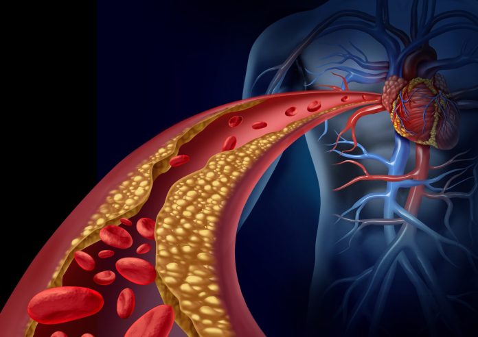
PDA is a serious medical condition involving atherosclerosis in the leg arteries and affects more than 200 million people worldwide.
These researchers were able to use a new magnetic-resonance imaging (MRI) technique at the end of exercise to understand the effects of PAD in the calves of patients with the disease and distinguish them from normal volunteers.
The approach they used, called chemical exchange saturation transfer, or CEST, produced results comparable to the current gold standard, which does not produce an image. CEST, they found, offered added benefits without requiring highly specialized equipment unavailable to many hospitals and researchers.
“The beauty of CEST is that it creates an image of energy stores in the muscle which we can match to images of blood flow,” said senior author Christopher M. Kramer, MD, chief of UVA Health’s Division of Cardiovascular Medicine and a professor of cardiology and radiology at the University of Virginia School of Medicine. “This gives us a new understanding of how atherosclerosis in the leg arteries causes problems in the muscles downstream.”
PAD affects more than 7% of Americans over age 40 and more than 29% of those over 70. The disease can cause pain when walking, coldness or numbness in the lower leg, painful leg or arm cramps, difficulty sleeping and erectile dysfunction, among other symptoms, though it also may cause no symptoms at all.
The lack of adequate blood flow to the limbs may make it difficult for wounds to heal and can, in severe cases, lead to amputation. Existing treatments include medicine to improve blood flow and manage pain; for appropriate cases, doctors may also consider options such as surgery or the placement of a stent to open clogged arteries.
To see if CEST would help improve treatment, the research team conducted a clinical trial comparing the technique with the current gold-standard approach, phosphorus magnetic resonance spectroscopy.
The researchers used CEST to image 35 volunteers with PAD and compared the results with imaging obtained from 29 control subjects after they performed calf exercise in the MRI scanner. They found that CEST was effective at identifying PAD in the lower legs, differentiating patients from normal subjects, and the results compared favorably to phosphorus magnetic resonance spectroscopy.
CEST, they concluded, could offer many advantages for researchers. CEST has better special resolution, creates an image and does not require costly equipment needed for phosphorus magnetic resonance spectroscopy. That means more centers could take advantage of the approach.
The technique can also be combined with other magnetic resonance imaging methods that measure blood flow in the calf to better understand the effects of PAD, the researchers note.
“Combining CEST with the techniques for measuring muscle blood flow with MRI enables an exciting new approach to studying potential benefits of established and novel therapies in this disease,” Kramer said.


