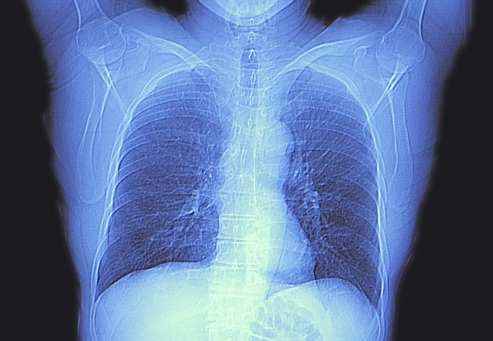
New research from the University of Texas MD Anderson Cancer Center says that colorectal cancer patients with certain clinical characteristics may benefit from more frequent screening via chest imaging to help identify and target cancer that has metastasized to the lungs.
While survival rates are improving due to improved screening rates, colorectal cancer (CRC) remains the third-leading cause of cancer deaths in the US. These cancers, when detected early, can be successfully treated and many CRC patients remain cancer-free for the rest of their lives. But, colon cancer spreads in up to 50% of all patients, with as many as 18% of patients developing metastatic cancer of the lungs. Finding lung nodules early improves survival rates, yet there are currently no published guidelines to help clinician know how often to screen CRC patients with chest CT or PET scans, or even which patients should be prioritized to receive such screenings.
“With this study, we sought to develop a strategy that is evidence-based to determine how frequently, at what intervals, and for how long patients at risk of developing lung metastases should undergo imaging of their chest,” said study co-author Mara Antonoff, MD, FACS, associate professor, thoracic and cardiovascular surgery at MD Anderson.
To help answer these questions, the team at MD Anderson turned to two databases at the cancer center that included both CRC cancer patients and thoracic cancer patients. The retrospective study grouped CRC patients by those who didn’t develop lung nodules and those who did and the timing of their diagnoses. The team then analyzed additional data of these patients to determine those most at risk of developing lung metastases.
Of the 1,600 patients with CRC in study, 233, or 14.6%, developed pulmonary metastases, with a median time of 15.4 months following colorectal surgery. The analysis of these data revealed a number of different risk factors for developing metastatic lung cancer among this group including age, neoadjuvant or adjuvant systemic therapy (such as chemotherapy or immunotherapy), lymph node ratio, lymphovascular and perineural invasion, and the presence of KRAS genetic mutations.
The analysis also showed that those patients most at risk of developing lung metastases shortly after surgery—within three months—had an elevated lymph node ratio, and a KRAS mutation.
“A concrete clinical application of this research, following validation, is to build evidence-based guidelines affecting chest surveillance in patients with resected colorectal cancer,” Nathaniel Deboever, MD, general surgery resident at the UTHealth Houston McGovern Medical School, and the lead author of the study “These guidelines will hopefully allow high-risk patients to undergo radiographic screening in a timely manner, permitting the early diagnosis of pulmonary disease.”
The team will now look to validate their findings using data from a different set of CRC patients with a goal of creating a set of clinically validate chest screening protocols that can be broadly adopted.
“There are many patients who receive cancer care outside of cancer hospitals, so having algorithms, pathways, and recommended protocols can be very helpful for providers who care for a lot of different diseases with rapidly changing recommendations,” Antonoff said. “I think this research is really just the tip of the iceberg.”


