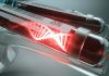






Researchers at Nagoya University, Japan, have developed a nanowire-based microfluidic device they say can improve the detection and analysis of cancer-associated extracellular vesicles (EVs) in blood samples. The new device uses zinc oxide nanowires modified with antibodies to selectively capture EVs linked to cancer while preserving both their surface proteins and internal microRNAs, which can then be analyzed. The findings, published in the journal Device, indicate that new device could potentially improve liquid biopsies for ovarian cancer as well as other cancers.
“In this study, we developed a nanowire microfluidic device capable of selectively capturing cancer-associated EVs with high efficiency, while suppressing nonspecific adsorption through simple chemical modification,” said senior author Takao Yasui, PhD, a professor in the graduate school of engineering at Nagoya University. “We also demonstrated that this approach maintains both EV membrane proteins and internal microRNAs intact, showing strong potential for highly sensitive analysis of cancer states.”
EVs are an emerging class of analytes because they carry molecular cargo such as messenger RNAs, microRNAs, and membrane proteins that reflect the cells from which they originate. To date, however, there have been challenges finding way to isolate EVs from complex biological fluids.
According to the researchers, techniques to isolate EVs such as ultracentrifugation, size-exclusion chromatography, and polymer-based precipitation can be time-consuming, require large sample volumes, and provide limited specificity. These methods also don’t reliably distinguish between EV subtypes.
Previously, investigators in the Yasui lab had developed zinc oxide nanowire devices capable of efficiently enriching EVs through charge interactions and hydrogen bonding. Further, the researchers noted that polyketone-coated zinc oxide nanowires improved the efficiency and purity of EV isolation within a microfluidic system designed for cancer diagnostics. Those findings led the team to explore whether polyketone chemistry could provide a more controlled method of attaching antibodies to the nanowires.
To do this, the team developed a platform based on N-hydroxysuccinimide ester-functionalized polyketones, known as pKNHS. They synthesized six pKNHS variants with different chain lengths and found that pKNHS 4.2 provided the most effective combination of nanowire stability and antibody immobilization.
The technology was first evaluated using cultured breast cancer cells. Antibody-free nanowires captured approximately 65% of CD9-positive EVs, while nanowires conjugated with CD9 antibodies captured efficiency of about 90%.
Next, the platform was testing using antibodies directed against ovarian cancer-associated markers CLDN3, FOLR1, and TROP2. These modified nanowires selectively recovered EVs from ovarian cancer cells and were subsequently used to isolate EVs from blood serum samples obtained from six patients with high-grade serous ovarian carcinoma and six individuals without cancer.
Analysis of the captured EVs showed distinct microRNA patterns of the EVs collected from cancer patients compared with those from the people without cancer. In total, the team identified 126 microRNAs shared among EVs captured using all three ovarian cancer markers. They also found unique microRNA populations linked to each marker, including 40 associated with CLDN3, 37 with FOLR1, and 45 with TROP2.
The system also facilitated the analysis of both EV membrane proteins and encapsulated microRNAs, which could scientists to better understand the relationships between surface markers and the associated molecular cargo.
Future work will focus on comparing the technology with existing clinical diagnostic methods and expanding its ability to capture additional EV subpopulations. The long-term goal is to apply the approach to noninvasive liquid biopsies and early diagnosis across a range of cancer types.