





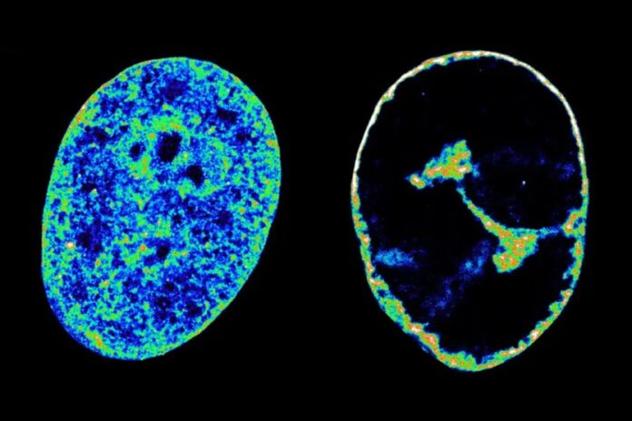
Researchers have discovered that the herpes simplex virus-1 (HSV-1) rearranges the three dimensional shape of the human genome to access genes it needs to replicate itself. For the first time, scientists have been able to study in detail how the virus rearranges the architecture of its host’s genome within hours of infection, opening the door for new therapeutic strategies that target this infection mechanism.
“HSV-1 is an opportunistic interior designer, reshaping the human genome with great precision and choosing which bits it comes into contact with,” said Esther González-Almela, PhD, associate professor at the Guangdong Provincial People’s Hospital (GDPH) in Guangzhou, China, and first author of the study. “It’s a novel mechanism of manipulation we didn’t know the virus had to exploit host resources.”
As a DNA virus, HSV-1 depends on the host cell’s molecular machinery to replicate itself. Previous research had shown that the virus takes over transcription of the human genome by hijacking the RNA polymerase II (RNAP II) enzyme, which is responsible for transcribing DNA into messenger RNA. However, it remained unknown how this process affected the three dimensional structure of the human genome.
“Interestingly, as the viral genes are being highly transcribed by RNAP II, the human genome undergoes a major shutoff,” write the researchers in Nature Communications. “Despite the efforts to elucidate the mechanisms that switch from human to virus expression, we are far from understanding how the changes in viral and cellular chromatin are mediated, and specifically, how the human genome is altered and manipulated by the virus. A potential limitation in previous studies has been the lack of single-cell analysis and high-resolution methods.”
The research team used single-molecule localization microscopy (SMLM) and genomic Hi-C techniques to visualize nanoscale changes to the chromatin structure in cells infected with HSV-1. Results showed that the virus recruited RNAP II and topoisomerase I (TOP1), an enzyme that snips DNA, away from their usual genome location and into virus replication compartments. This causes transcription to collapse across the whole genome and results in chromatin compacting into a dense shell 30% of its original volume within three hours from infection.
The researchers noted that this infection strategy is very different from that of RNA viruses, such as influenza or SARS-CoV-2, opening new possibilities to target vulnerabilities unique to the herpes virus. Their findings showed that inhibiting TOP1 was enough to completely block the replication of the virus, presenting the inhibition of topoisomerase enzymes as a promising antiviral strategy to explore in future work.
“In cell culture, inhibiting this enzyme stopped the infection before the virus could make a single new particle,” said Maria Pia Cosma, PhD, senior author of the study and professor at GDPH and the Centre for Genomic Regulation (CRG) in Barcelona, Spain. “That gives us a potential new therapeutic target to stop infection.”