





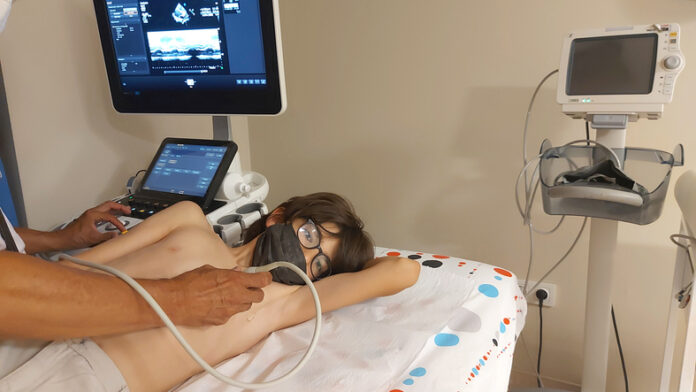
A novel AI model that mimics a human reader to assess multiple key measures of cardiac structure and function on echos could help identify heart abnormalities in areas where expert resources are scarce.
The PanEcho multitask deep learning system could act as a preliminary reader to identify missed abnormalities on echocardiograms, one of the pillars of cardiovascular diagnostics.
The tool described in JAMA, could act as an efficient screening system for point-of-care examinations by nonexperts and also support expert readers by identifying key abnormalities for further interrogation. The model and source code have been publicly released to accelerate research on AI-enabled echocardiographic interpretation.
“This AI system was developed to address a critical gap in AI for echocardiography: the predominance of single-view, single-task models,” explained the researchers, led by Gregory Holste, a PhD candidate from the University of Texas at Austin.
These single-task models pose considerable challenges in terms of implementation, especially for situations such as deploying a handheld device for point-of-care use. PanEcho offers a better perspective by performing end-to-end estimation of all variables that form the core of a complete echocardiographic report.
“This unified approach is closer to clinical practice, in which a cardiologist synthesizes multiple views and imaging modes to assess all aspects of cardiovascular health, and more efficiently scales to clinical deployment,” the team noted.
More than 7.5 million echocardiographic studies are performed annually in the U.S. alone, and increasing referrals contribute to rising health care costs, with accurate reporting requiring time, skilled acquisition, and expert readers.
To address these challenges, PanEcho was developed and validated in more than a million echo videos, spanning a broad range of acquisitions and phenotypes. It uses a view-agnostic deep learning model, which automates their interpretation and performs 39 diverse interpretation tasks.
The system automatically interprets key aspects of echocardiograms that range from ventricular structure and function to valvular disease and vascular structure and function, while maintaining accuracy across different geographic locations and time periods.
It improves on the current state of AI-enabled echocardiography, enabling simultaneous estimation of all key parameters of cardiac structure and function from any combination of 2D views.
The system was developed using transthoracic echocardiography (TTE) studies conducted at Yale New Haven Health System (YNHHS) hospitals and clinics from January 2016 to June 2022 during routine care.
It was then internally validated in a later YNHHS cohort from July to December 2022 before being validated across four diverse external cohorts, with the AI predictions compared with assessment of the interpreting cardiologist. Together, the study included 1.2 million echocardiographic videos from 32, 265 TTE studies of 24,405 patients across YNHHS hospitals and clinics.
The AI system performed 18 diagnostic classification tasks with a median area under the receiver operating characteristic curve (AUC) of 0.91 and estimated 21 echocardiographic parameters with a median normalized mean absolute error of 0.13 in internal validation. The model accurately estimated left ventricular ejection fraction, and detected moderate or worse left ventricular systolic dysfunction, right ventricular systolic dysfunction, and severe aortic stenosis.
The AI system maintained excellent performance in limited imaging protocols, the researchers report, performing 15 diagnosis tasks with a median AUC of 0.91 in an abbreviated TTE cohort and 14 tasks with a median AUC of 0.85 on real-world point-of-care ultrasonography acquisitions from YNHHS emergency departments.
“The model is open-source, extensively validated internationally, and at the point of care and may signal a shift from specialized single-task models toward generalized multitask AI models poised for clinical deployment,” the authors concluded.