




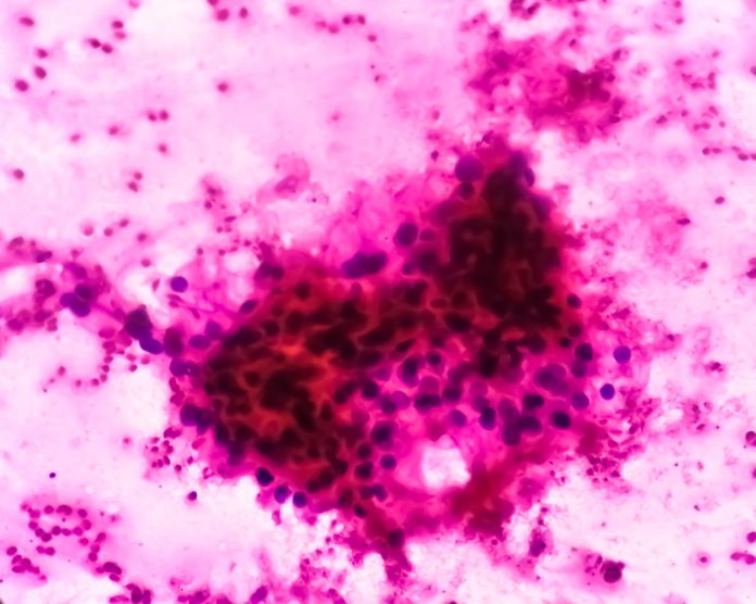
Giant-nucleus cells’ response to iron could be a harbinger of otherwise undetectable cancer, according to a research team at Nagoya University. This team’s study of kidney cancer in rats suggests giant-nucleus cells that survive iron-induced damage may form the foundation of early-stage cancer. While this study was only in kidney cells, the team sees this finding possibly applying to other cancers as well.
The team proposes these cells, which are easy to identify under a microscope, could be good biomarkers of cancer. The study was published in the journal Redox Biology, and the lead author is Shinya Toyokuni, MD, PhD, professor emeritus at Nagoya University Graduate School of Medicine.
“The association between abnormal nuclear morphology and the molecular programs identified in our rat kidney model was also detectable in human breast epithelial tissue, supporting the idea that a BRCA1-associated karyomegalic state may not be restricted to the kidney,” Toyokuni told Inside Precision Medicine.
The study used two groups: Wild-type rats and some with a single mutated BRCA1 gene. The rats were administered ferric nitrilotriacetate, a carcinogenic iron compound that causes kidney damage. The team then examined kidney tissue samples from them across various time points. The morphological analysis included seven patients carrying germline BRCA1 mutations and 15 non-carriers. In total, they examined 7,662 ductal epithelial nuclei: 2,141 nuclei from BRCA1 mutation carriers and 5,521 from non-carriers.
They validated their findings by analyzing additional animals and comparing the results with human cancer datasets. Using spatial transcriptomics, they mapped gene activity in individual cells and conducted computational analyses to quantify the size, shape, and density of cell nuclei in those samples.
The team saw giant-nucleus cells emerge within one week of ferric nitrilotriacetate administration. The cells increased expression of cancer-related genes such as Myc and Met, and developed resistance to ferroptosis—an iron-dependent form of cell death.
BRCA1-mutant rats developed giant-nucleus cells with distinct biological profiles from wild-type rats, suggesting BRCA1 deficiency impairs DNA repair, allowing more precancerous giant-nucleus cells to survive. Even morphologically normal cells near giant-nucleus cells exhibited gene expression changes that might promote their survival.
“At the individual nucleus level, ductal epithelial nuclei from BRCA1 mutation carriers showed significant differences in eccentricity, solidity and extent, indicating less compact and more irregular nuclear contours,” Toyokuni said. “More importantly, within the BRCA1-mutant group, the activity of the cancer-associated Karyo2 transcriptional program correlated strongly with nuclear eccentricity, while the shared KaryoCore program correlated with nuclear solidity. These relationships were not observed in the non-carrier group.”
Through analysis of gene activity and nuclear morphology, the researchers classified giant-nucleus cells into six types. One type showed extremely large nuclei and signs of stress-induced growth arrest, consistent with profiles from previous studies. Another showed increased cancer-related gene activity and elongated nuclei, pointing to a potential precancerous state.
Through additional electron microscopy and cellular respiration measurements, the team confirmed that cells from BRCA1-mutant rats exhibited mitochondrial remodeling, which led to impaired iron handling and respiratory function.
The researchers also analyzed data from the TCGA-KIRC, a large public database of kidney cancer patients. They found that patients whose tumors shared gene patterns with cancer-prone giant-nucleus cells had shorter survival times, while those with patterns seen in healthier giant-nucleus cells had better outcomes. These results suggest that not all giant-nucleus cells carry the same risk.
In a small pilot study, the researchers also observed similar abnormal nuclear features in breast tissue from seven individuals with inherited BRCA1 mutations, compared to 15 without the mutation.
“Using spatial transcriptomics, we categorized giant-nucleus cells into six types for the first time and detailed their cancer-linked traits,” Toyokuni said. “By comparing these features with human data, we found that these cell types could help predict patient outcomes.”
Outstanding questions are: how do giant-nucleus cells develop into tumor cells, whether suppressing mitochondrial changes or ferroptosis resistance can prevent cancer, and whether nuclear shape and gene patterns can help diagnose or predict kidney cancer in larger patient groups.