





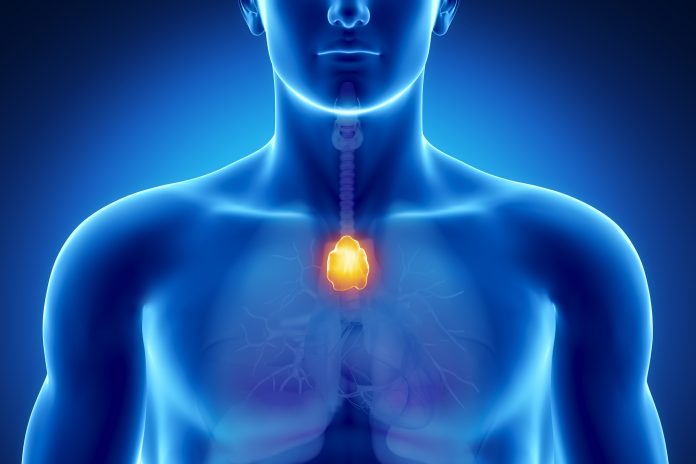
Research led by Mass General Brigham has shown that patients who receive a high incidental dose of radiotherapy (RT) to the thymus during treatment for non-small-cell lung cancer (NSCLC) have an increased risk for metastasis and death, especially if they have preserved thymic function.
The study “establishes the clinical importance of unintended thymic irradiation in patients with NSCLC undergoing thoracic RT,” and highlights a need for a “paradigm shift in thoracic radiation planning, where immunological health is prioritized alongside traditional oncological targets and organs at risk,” write first author Vasco Prudente, from the Artificial Intelligence in Medicine (AIM) Program at Mass General Brigham & Harvard Medical School, and colleagues in the Annals of Oncology.
The researchers explain that the thymus, which is responsible for producing and educating T cells, has traditionally been considered to have limited functional importance in adulthood. This long-held view has meant that the organ has not been included in adult oncology treatment planning, even though immune function strongly influences outcomes in people with cancer.
However, recent work by the AIM team showed that many adults retain measurable thymic function throughout life, and that healthier thymuses are associated with lower mortality, lower risk of cardiovascular disease and cancer, and improved responses to modern immunotherapies.
Because RT in patients with NSCLC may incidentally expose the thymus, located behind the breastbone, to radiation, the researchers hypothesized that thymic radiation dose may be negatively associated with clinical outcomes.
They investigated their theory using data from 1107 patients with locally advanced NSCLC from the Phase III RTOG-0617 trial (n=460), and two real-world patient cohorts (HARVARD-CRT, n=422 and HARVARD-DURVA, n=225).
They found that the mean thymic dose was 46.1 Gy in RTOG-0617, 49.4 Gy in HARVARD-CRT, and 32.0 Gy in HARVARD-DURVA. Most (92%) patients received concurrent chemotherapy and the median time to distant metastases across all patients was 0.9 years, with a median overall survival of 1.8 years.
After adjusting for known prognostic variables, including radiation dose to the heart, lungs, and esophagus, tumor volume, cancer stage, smoking status, and the interaction between mean thymic dose and baseline thymic health, the investigators observed that a 1-Gy increase in mean thymic dose was associated with significant 29%, 33%, and 95% increased risks for distant metastases in the RTOG-0617, HARVARD-CRT, and HARVARD-DURVA study participants, respectively.
In an exploratory analysis, Prudente and team showed that patients who received a mean thymic dose that exceeded a potential clinically meaningful threshold of 35 Gy had a significant 40% greater risk for distant metastasis than those below this threshold.
The association was attenuated, however, when they stratified the cohort by thymic health, which was assessed using an AI model that analyzes radiographic features on chest computed tomography scans and quantifies thymic function on a scale from 0 (minimal thymic health) to 1 (maximal thymic health).
Further analysis showed that higher mean thymic dose was associated with an increased risk for metastases and death in patients with preserved thymic health but not in those with impaired thymic function.
“I think this is one of the exciting aspects of the work: in principle, thymic health could become another piece of information available when treatment is being planned,” co-senior author Hugo Aerts, PhD, director of the AIM Program at Mass General Brigham told Inside Precision Medicine.
He added that although patients with preserved thymic health before treatment generally had the best outcomes overall, within this group, higher radiation doses to the thymus were associated with a substantially greater risk for distant metastasis.
“This raises the possibility of a new form of precision medicine: rather than applying the same radiation constraints to every patient, we might eventually use an individual’s thymic health to identify who has functioning immune tissue worth protecting and personalize the radiation plan accordingly,” said Aerts. “This could be particularly relevant as immunotherapy becomes increasingly integrated with radiation. Our previous work showed that patients with healthier thymuses tend to respond better to immunotherapy. Taken together, these findings suggest that understanding and preserving a patient’s immune health may become an important part of personalizing cancer treatment.”
The researchers also showed that declines in thymic health were dependent on radiation dose and that RT planning could feasibly be reoptimized to spare the thymus without compromising tumor coverage or cardiopulmonary constraints.
“This would not require a new treatment or new technology,” noted Aerts. “Thymus-sparing could therefore potentially be incorporated into routine practice relatively quickly.”
The researchers are now planning to validate their findings in prospective studies. “We want to determine whether deliberately reducing radiation exposure to the thymus preserves thymic and immune function and, ultimately, whether this translates into fewer metastases and better survival,” said Aerts.
The group is also studying the impact of thymic health across other cancers and treatments.