





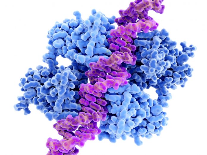
Mutations in the tumor suppressor TP53 are among the most common genetic alterations in cancer, making mutant p53 an attractive target for personalized immunotherapies. However, new research suggests that identifying a tumor mutation may be only the first step toward finding an effective T-cell target.
Researchers led by Dana-Farber Cancer Institute and Harvard Medical School found that tumors can actively restrict the presentation of p53-derived neoantigens on their surface, allowing cancer cells to evade T-cell recognition. The findings, published in Immunity, also identify antigen-processing mechanisms that could potentially be manipulated to make tumors more visible to the immune system.
Predicted neoantigens do not always reach the cell surface
T cells recognize short peptides presented by human leukocyte antigen (HLA) molecules rather than directly reading mutations in tumor DNA. Consequently, a mutation predicted to produce a strong neoantigen is therapeutically useful only if the resulting peptide is processed, loaded onto an HLA molecule and displayed at sufficient levels on the tumor surface.
The researchers used an ultrasensitive mass spectrometry-based immunopeptidomics platform alongside tumor genomics, structural biology, and engineered T cells to directly investigate p53 peptide presentation.
The discrepancy between prediction and presentation was substantial. Among 175 predicted wild-type p53 peptide candidates across relevant HLA molecules, only five were robustly detected. Several common p53 hotspot mutations also occurred in regions that produced no detectable surface targets.
The results suggest that genomic sequencing and computational HLA-binding predictions alone may overestimate the pool of actionable neoantigens, emphasizing the value of experimentally confirming peptide presentation when selecting targets for precision T-cell therapies.
ERAP1 provides a route for immune escape
The researchers identified several mechanisms through which tumors could lose otherwise promising targets.
For the p53 I195F mutation, they found that endoplasmic reticulum aminopeptidase 1 (ERAP1), an enzyme involved in trimming peptides before HLA presentation, could destroy the neoantigen before it reached the cell surface. Knocking out ERAP1 or pharmacologically inhibiting the enzyme restored recognition of tumor cells by p53-specific T cells in laboratory experiments.
A different limitation emerged for the commonly studied p53 R175H neoantigen. Although researchers generated highly sensitive T-cell receptors against the target, the mutant peptide bound weakly to HLA-A*02:01, resulting in an unstable, short-lived peptide-HLA complex and reduced T-cell activation and tumor-cell killing.
Expanding the precision immunotherapy toolkit
The findings support a therapeutic concept the researchers describe as “immunopeptidome shifting,” pharmacologically altering the peptides that tumors display rather than focusing exclusively on strengthening the immune response.
Potential approaches include ERAP1 inhibitors, compounds that alter peptide binding to HLA molecules and drugs that modify RNA splicing. Such strategies could potentially expose new tumor-specific targets and improve responses to checkpoint inhibitors, engineered T-cell therapies or other antigen-directed treatments.
The work remains preclinical and does not demonstrate that manipulating antigen presentation can convert immunologically “cold” tumors into responsive tumors in patients. However, it highlights an increasingly important consideration for precision immunotherapy: the presence of a cancer mutation does not necessarily mean the immune system can see it.