Maltreatment of children prior to adolescence, which includes physical and emotional abuse, has been shown to adversely affect healthy brain development. Adults who were abused as young children often show atypical brain structures and resultant psychiatric disorders.
But neuroscientists have also shown that during adolescence—and shortly thereafter—the neocortical regions of the brain undergo a major reorganization, which provides a window to treat some of these disorders. But the question of whether there is an identifiable biological mechanism that can be targeted during this time to treat some of these disorders has remained in question.
Now, new research from investigators in Japan and U.S. have pinpointed a specific hormone and the mechanism by which is created that might altered as a result of maltreatment, providing a potential pathway for treatment.
The study, published in Translational Psychiatry, was led by Shota Nishitani, PhD, and Akemi Tomoda, MD, PhD, from the University of Fukui, Japan, and Alicia K. Smit, PhD, from Emory University.
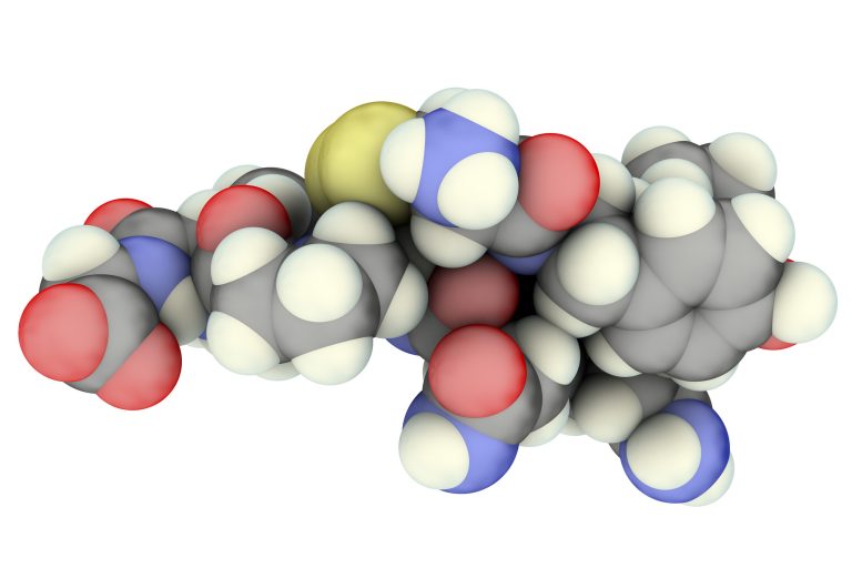
The hormone in question is oxytocin, which plays a key role in the capacity to feel empathy and love, and learn prosocial behaviors like bonding. The biological mechanism that the study focused on is DNA methylation, which regulates the secretion of oxytocin.
Based on insights from previous studies, which showed that methylation of the oxytocin gene was associated with both brain structure and social behaviors, the researchers hypothesized that child maltreatment could be related to the oxytocin gene methylation levels and, in turn, altered brain structures and brain development during childhood and adolescence. They put this hypothesis to the test through various experiments involving DNA sampling and multi-mode brain imaging of children who had been maltreated and compared their outcomes against of children who did not suffer from maltreatment.
“Child maltreatment dysregulates the brain’s oxytocinergic system, resulting in dysfunctional attachment patterns. However, how the oxytocinergic system in children who are maltreated (CM) is epigenetically affected remains unknown. We assessed differences in salivary DNA methylation of the gene encoding oxytocin (OXT) between CM (n = 24) and non-CM (n = 31), alongside its impact on brain structures and functions using multi-modal brain imaging (voxel-based morphometry, diffusion tensor imaging, and task and resting-state functional magnetic resonance imaging),” write the investigators.
“We found that CM showed higher promoter methylation than non-CM, and nine CpG sites were observed to be correlated with each other and grouped into one index (OXTmi). OXTmi was significantly negatively correlated with gray matter volume (GMV) in the left superior parietal lobule (SPL), and with right putamen activation during a rewarding task, but not with white matter structures.
“Using a random forest regression model, we investigated the sensitive period and type of maltreatment that contributed the most to OXTmi in CM, revealing that they were 5–8 years of age and physical abuse (PA), respectively. However, the presence of PA (PA+) was meant to reflect more severe cases, such as prolonged exposure to multiple types of abuse, than the absence of PA. PA+ was associated with significantly greater functional connectivity between the right putamen set as the seed and the left SPL and the left cerebellum exterior.
“The results suggest that OXT promoter hypermethylation may lead to the atypical development of reward and visual association structures and functions, thereby potentially worsening clinical aspects raised by traumatic experiences.”
The DNA tests showed that a specific region of the oxytocin gene was consistently more methylated in maltreated children. The researchers then linked this hypermethylation with changes in brain structure and activity.
“We found a higher DNA methylation rate to be associated with a smaller volume of the left superior parietal lobe in the dorsal attention network, which is important for the coordination of one’s eye movement with the recognition of other’s gaze,” explains Tomoda. “We also noted low brain activity in the right putamen, which is a part of the reward system network.”
Additionally, the researchers determined through statistical analyses that children who were maltreated showed higher levels of DNA methylation than children of the same age in the general population. When DNA methylation was high, there was a decrease in volume in the brain attention network and a decrease in activity in the reward system network. The DNA methylation was also associated with a specific period (58 years old) and higher levels of maltreatment such as physical abuse, especially among children who had received maltreatment.
The team says this is the first study to examine the relationship between methylation in the oxytocin gene and child maltreatment, and the insights could have groundbreaking clinical applications.
“The reversible nature of the DNA methylation implies that it may be possible to develop methods to de-methylate the oxytocin gene region. If we can demonstrate that such methods could reverse brain function alterations and trauma symptoms in maltreated children, we could create entirely new therapies to improve their quality of life,” notes Smith.


