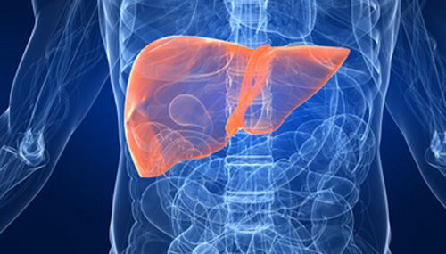
Hepatocellular carcinoma (HCC) is the most common type of liver cancer with incidence and mortality rates rising globally. Often caused by previous viral infections from hepatitis B and hepatitis C, HCC detection early in the course of disease remains less than optimal.
To improve early diagnosis, researchers from the National Cancer Institute (NCI) developed a new HCC screening blood test using a synthetic human virome. The VirScan test probes for a viral signature of past exposure (VES).
In a study published in Cell, NCI researchers found that the screening tool identified cancer patients prior to a clinical diagnosis.
“We hypothesize that unique viral exposure signatures (VESs) resulting from virus-host interactions could reflect a cascade of events that may alter the risk of developing HCC,” the authors write. “Such signatures may serve as early detection biomarkers and offer knowledge about potentially modifiable factors for early onset of HCC.”
Viral Exposure Signature
The team relied on VirScan, is a phage immuno- precipitation sequencing (PhIP-seq) technology, to detect the exposure history to all known human viruses. VirScan applies a phage display library that covers 93,904 viral epitopes, representing 206 human viral species and over 1,000 viral strains, to screen for previous exposure history.
To discover a VES related to HCC, the researchers gathered blood profiles from 899 people, including 150 people with HCC, from a NCI-UMD (National Cancer Institute-University of Maryland) case-control study of liver cancer. A phage particle with an epitope that was recognized by a participant’s antibody was immunoprecipitated and the encoding DNA barcode was then sequenced.
After sorting the data from these 1,000 + patients, they ultimately identified a viral exposure signature that reflected differences between individuals with HCC compared with people with chronic liver disease and healthy volunteers. This signature contained evidence from 61 different viruses.
Validating the results
They validated the results in a study of 173 at-risk individuals with chronic liver disease who had been followed for HCC development for 20 years. These individuals were part of a prospective NIDDK (National Institute of Diabetes and Digestive and Kidney Diseases) at-risk cohort for HCC.
During that time, 44 of the participants developed HCC. Using blood samples taken when the cancer was diagnosed, the signature correctly identified all of those who developed HCC (area under the curve AUC=0.98).
Perhaps of greatest importance, the screen also detected HCC in their blood samples taken at the start of the study – in some cases up to ten years prior to their HCC diagnosis (AUC=91).
The AUC is a measure of the accuracy of a quantitative diagnostic test. An AUC of 0.5 indicates that a test is no better than chance in identifying disease, whereas an AUC of 1.0 represents a test with perfect accuracy.
When tested against alpha-fetoprotein (AFP), the current blood analyte used in screening HCC methods currently, the viral exposure signature tool more accurately detected the presence of HCC. The AUC for AFP testing was 0.62 for AFP
Bi-annual screening with ultrasound and/or an AFP blood test are the current methods for screening people with known HCC risk factors. However, most HCC cancers are found when the disease is already metastatic or incurable. Like all cancers, early HCC detection raises the chances of successful treatment.
“We need a better way to identify people who have the highest risk for HCC and who should get screened more frequently,” said lead author Xin Wei Wang, PhD, co-leader of the NCI Center for Cancer Research Liver Cancer Program. “We established a viral exposure signature that can predict HCC among at-risk patients prior to a clinical diagnosis, which may be useful in HCC surveillance.”


