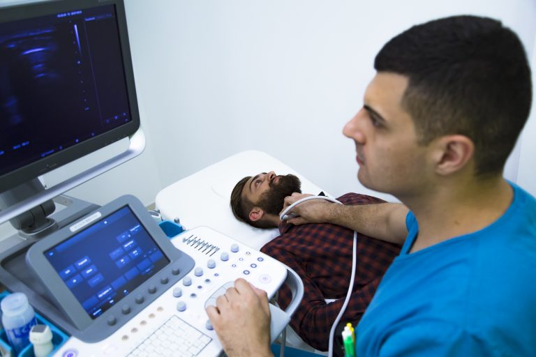
Researchers at Harvard have developed an ultrasound-image based artificial intelligence (AI) driven platform that can help detect thyroid cancer.
While recent developments in image recognition using AI are being used effectively to scan a number of different medical images, such as histology slides used for cancer diagnosis, ultrasound poses some difficulties.
Unlike other imaging machines and techniques used for diagnostic purposes, such as magnetic resonance imaging or computed tomography, as well as other high quality still images, the quality of ultrasound imaging depends a lot on the operator, their experience and the patient’s body.
To try and overcome some of these potential image difficulties, the researchers combined four different AI methods, namely, radiomics—analyzing different data points on the images, topological data analysis—evaluation of spatial relationships between points on the images, and a combination of deep learning and machine learning. Deep learning networks are made up of a complex set of algorithms modeled on the human brain and machine learning uses algorithms to perform tasks and learn from them.
“By integrating different AI methods, we were able to capture more data while minimizing noise. This allows us to achieve a high level of accuracy in making predictions,” said Annie Chan, Director of the Head and Neck Radiation Oncology Research Program at the Mass General Cancer Center, who is presenting this work this week at the 2022 Multidisciplinary Head and Neck Cancers Symposium in Arizona.
To test and validate their model, Chan and colleagues used diagnostic data from needle biopsies and high-quality ultrasound images taken of patients with suspected thyroid cancer. Overall, 464 patients and 511 nodules were tested. There were three training sets for the model an internal training set (103 malignant, 259 benign nodules), an internal validation set (51 malignant and 98 benign nodules) and an external validation set (270 malignant and 50 benign nodules).
Combining all four techniques allowed the researchers to achieve a predictive accuracy with the model of 98.7% on the internal dataset. On external validation this model was 93% accurate at predicting malignancy. In comparison, using any of these techniques alone instead of in combination reduced predictive accuracy to 80–89%.
Notably, the combined model was also able to distinguish pathological stage of the tumors with good accuracy (89–98%) and also identify those with the BRAF V600E mutation subtype of thyroid cancer, which responds well to targeted therapy.
“Thyroid cancer is one of the most rapidly increasing cancers in the United States, largely due to increased detection and improved diagnostics. We have developed an AI platform that would examine ultrasound images and predict with high accuracy whether a potentially problematic thyroid nodule is, in fact, cancerous. If it is cancerous, we can further predict the tumor stage, the nodal stage and the presence or absence of BRAF mutation,” said Chan in a press statement. “If caught early, this disease is highly treatable, and patients generally can expect to live a long time after treatment.”


