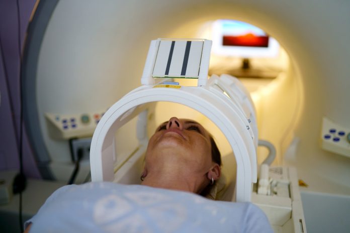
A cheaper, safer, ultra-low energy magnetic resonance imaging (MRI) scanner has been developed that has the potential to become a powerful point-of-care tool, especially in low-to middle-income countries.
The technology has the potential to offer accessible and affordable screening and could one day be used for diagnostics, risk assessment and treatment decisions in many diseases, such as stroke.
The ultra-low field MRI machine, outlined in the journal Science, does not need radiofrequency or magnetic shielding cages, uses a standard wall power outlet, and costs a fraction of current clinical scanners.
It uses a magnetic strength of just 0.05 tesla versus the 1.5 to 3 tesla usually seen with current versions in the clinic, with data-driven deep learning to enhance the image quality and reduce scan time.
Yujiao Zhao, PhD, from the University of Hong Kong, and co-workers note that clinical MRI procedures remain mostly unattainable for two-thirds of the world’s population.
“This scanner is compact and potentially mobile, and can be manufactured, maintained, and operated at a low cost,” they report.
MRI offers near-limitless opportunity for visualizing tissue and identifying pathology and is routinely used to diagnose and select treatments for brain, heart, cancer, and orthopedic conditions.
Images are created by taking advantage of the magnetic fields generated by the spin of hydrogen atoms.
The spins are manipulated by radio-frequency pulses that generate contrasting signals in pathology and normal tissue, and a constant powerful magnetic field is applied to align most protons in the body to help differentiate the two.
Most current clinical MRI scanners use superconducting magnets to generate a constant adequate magnetic field, which require liquid helium to cool them and use high quantities of energy.
But the ultra-low-field machine in the current study enables whole-body imaging by placing two permanent magnet plates above and below the body in an open configuration. Using noise-sensing coils, optimized scan protocols and AI-based image reconstruction, the machine was able to produce image qualities comparable to those produced with 3 tesla.
The team tested the machine on healthy volunteers and were able to capture the brain, spine, abdomen, lung, musculoskeletal and cardiac images.
Deep learning image formation was able to substantially augment the 0.05 tesla image quality by exploiting computing and extensive high-field MRI data, the team notes.
Spinal images showed intervertebral disks, the spinal cord, and cerebrospinal fluid.
Abdominal images showed major structures such as the liver, kidneys and spleen, while lung images showed pulmonary vessels and parenchyma.
Knee images included cartilage and meniscus, while cardiac cine images showed left ventricle contraction and neck angiography displayed carotid arteries.
In an accompanying perspective article, Udunna Anazodo, PhD, from McGill University, and Stefan du Plessis, from Stellenbosch University, say that low-field MRI has “great potential” but that there are several challenges ahead.
These include limited experience in its clinical use, the need for radiologist retraining, and AI solutions that require big data.
“Low-field MRI has yet to mature to enable cost-effective access to medical imaging,” they write.
“Its potential as an essential and environmentally sustainable health technology will be proven when many communities around the world can use low-field MRI without barriers.”


