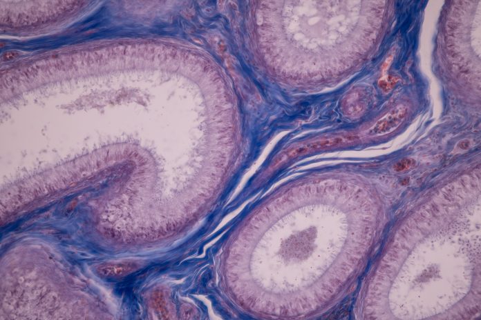
Research led by the Wellcome Sanger Institute near Cambridge has mapped the cells involved in the development of the ovaries and testes, which controls whether someone will become male or female.
The work forms part of the Human Cell Atlas project, an initiative to map all the cells in the human body that has already covered a number of other organs.
In this study, published in Nature, the researchers analyzed around 500,000 cells from human gonadal tissue sampled at regular intervals between week six and 21 of pregnancy using single cell sequencing and spatial transcriptomics. They also created a similar map in mice.
The study gave the researchers a better insight into how the cells interact with each other and are arranged during their journey to becoming a testis or an ovary and showed significant differences between humans and mice.
The team identified the cell type that is the first to express the sex determining gene, which they named Early Supporting Gonadal Cells. These cells are found in both mice and humans, peaking around 6 weeks after conception, but the gene expression associated with these cells differs between the two species. For example, in humans these express stem-cell markers such as TSPAN8 and LGR5.
“Gonadal development is a complex process and it is only at single-cell resolution that you start to see all of the cell types involved in sex differentiation and can narrow down the timeframe in which this process occurs,” commented first author Luz Garcia-Alonso, a researcher at the Wellcome Sanger Institute, in a press statement.
“The characterization of Early Supporting Gonadal Cells and their role in kick-starting sex determination is an exciting discovery that will help researchers to better understand this crucial time in human development.”
They researchers also found human SIGLEC15+ and TREM2+ macrophages in the developing testes that were similar to those seen in the brain and bone.
“It was fascinating to find macrophage populations in the gonads that we’re used to seeing in other organs, considering the very different purposes of the testes, the brain and bone. However, drawing parallels between the immune requirements of each organ can help us to understand the role of these macrophage populations wherever they occur in the body. In terms of evolution, if you have a biological function that could be useful elsewhere, why not use it,” said Valentina Lorenzi, a co-first author of the paper also from the Wellcome Sanger Institute.
The scientists also hope their findings will help to better understand the physiology of people born with differences in sex development that can occur in the womb and often result in atypical anatomy. The new information could also help to understand and treat different causes of infertility better.


