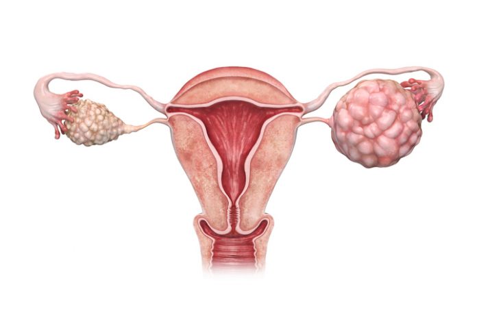
New findings from researchers at Nagoya University in Japan have revealed new potential biomarkers for ovarian cancer found in extracellular vesicles (EVs). The three previously unknown membrane proteins in extracellular vesicles (EVs) derived from high-grade serous ovarian carcinoma (HGSOC), a deadly subtype of epithelial ovarian cancer (EOC).
The group used a unique technology consisting of polyketone-coated nanowires (pNWs), tp capture the EV proteins, demonstrating the clinical utility of their detection method and the identification of potential biomarkers for this type of ovarian cancer.
In their research, published this week in the journal Science Advances, the team noted that “In summary, the HGSOC-specific marker detection by pNW are a promising platform as clinical biomarkers, and these insights provide detailed proteomic aspects of diverse EVs in HGSOC patients.”
EOC is a highly lethal gynecological cancer, and ranks as the seventh most common cause of cancer deaths among women worldwide, the authors noted. And because of the difficulty of early-stage screening, most cases are diagnosed at an advanced stage, at which point the five-year survival rate is less than 45%. Of the different subtypes of EOC, HGSOC accounts for about 75% of cases, with a fatality rate of nearly 90%. “There has been no specific and sensitive biomarker for HGSOC, and CA125, which is most commonly used in clinical diagnosis, has proven not to be effective for early detection of ovarian cancer,” the investigators stated.
The discovery of new biomarkers for ovarian cancer is important as the disease is difficult to identify during its early stages when it could most easily be treated. One approach to detecting cancer is to look for EVs, and especially small exosome proteins released by the tumor cells, the authors noted. “Cancer cell–derived EVs have unique protein profiles, making them promising targets as potential disease biomarkers.”
As these proteins are found outside the cancer cell, they can be isolated from body fluids, such as blood, urine, and saliva. However, EVs in body fluids are quite heterogeneous, the team pointed out, and use of such potentital biomarkers is hindered by the lack of reliable ones for the detection of ovarian cancer, the scientists stated. “HGSOC-specific EV protein markers with practical utility have not been identified … as rigorous assessments for EV proteomics have been a major challenge in the field, disease-specific EV markers are still widely lacking.”
For their reported study, the researchers extracted both small and medium/large EVs from HGSC, and analyzed them using liquid chromatography-mass spectrometry to analyze the proteins. “First, we investigated the size distribution of EVs and found that small EVs (sEVs) were better targets as biomarkers than medium/large EVs (m/lEVs),” the authors stated. “We then performed shotgun proteomics by using liquid chromatography–tandem mass spectrometry (LC-MS/MS) to reveal unique protein profiles for each EV size class with substantial functional differences between small and large EVs.”
The research did pose some challenges. “The validation steps for the identified proteins were tough because we had to try a lot of antibodies before we found a good target,” said Yokoi. “As a result, it became clear that the small and medium/large EVs are loaded with clearly different molecules.”
Continued analyses then highlighted individual proteins that might represent potential ovarian cancer biomarkers. “Further investigation revealed that small EVs are more suitable biomarkers than the medium and large type,” Yokoi added. “We identified the membrane proteins FRα, Claudin-3, and TACSTD2 in the small EVs associated with HGSC.”
Having identified the candidate biomarker proteins, the team then set out to determine whether they could capture EVs in a way that would allow them to identify the presence of cancer. To do this, nanowire specialist Takao Yasui, PhD, of the Graduate School of Engineering at Nagoya University, combined his research with that of Yasuhide Inokuma, PhD, at the Japan Science and Technology Agency, to create polyketone chain-coated nanowires (pNWs). This technology was ideal for separating exosomes from blood samples.
“pNW creation was tough,” Yokoi said. “We must have tried 3–4 different coatings on the nanowires. Although polyketones are a completely new material to use to coat this type of nanowire, in the end, they were such a good fit.”
To test the clinical utility of the pNW platform, the researchers used it to extract sEVs from samples of ascitic fluid from HGSOC patients, and from noncancer patients. In EOC, ascites is a major abnormal body fluid that accumulates in the peritoneal cavity in almost all cases, and especially in HGSOC patients, they explained. The ascites sEVs were then further analyzed. “Ascites sEVs, purified using pNW, were subjected to the single-particle imaging measurement assay (ExoView).” The results confirmed that sEVs positive for FRα, Claudin-3, or TACSTD2, were present in high concentrations in many HGSOC patients. Concluding on their findings, the team wrote, “… our pNW-ExoView platform revealed the specific elevated presence of FRα/Claudin-3/TACSTD2 on sEVs harvested from HGSOC patient ascites that can be useful HGSOC biomarkers.”
Yokoi added, “Our findings showed that each of the three identified proteins is useful as a biomarker for HGSCs. The results of this research suggest that these diagnostic biomarkers can be used as predictive markers for specific therapies. Our results allow doctors to optimize their therapeutic strategy for ovarian cancer, therefore, they may be useful for realizing personalized medicine.”
In their report, the authors concluded: “The pNW platform is well-suited for clinical application and may contribute to making EV-based diagnostics straightforward. There is broad potential for pNW-based applications for isolating further specific EVs in circulating body fluids.”


