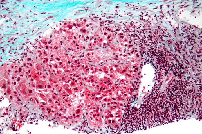
Researchers are looking to improve immunotherapy for liver cancer or hepatoceulluar carcinoma (HCC). Although immunotherapy has become the standard therapeutic method for fighting HCC, its response rate has only been approximately 30% in patients with advanced disease. Now, researchers at Osaka University report they have developed an analytical method that could help predict if an HCC patient would successfully respond to immunotherapy.
The findings, “Multiomics identifies the link between intratumor steatosis and the exhausted tumor immune microenvironment in hepatocellular carcinoma,” are published in the journal Hepatology.
“Immunotherapy has become the standard-of-care treatment for HCC, but its efficacy remains limited,” wrote the researchers. “To identify immunotherapy-susceptible HCC, we profiled the molecular abnormalities and tumor immune microenvironment (TIME) of rapidly increasing nonviral HCC.”
TIME is the area inside the tumor in which various immune cells and other cell types interact and send/receive messages and signals.
“We examined 113 patients that developed HCC without hepatitis virus infection,” said Hiroki Murai, lead author of the study. “We used a multi-omics approach, meaning we examined the DNA sequences of specific genes frequently mutated in HCC, but also performed a more general assessment of RNA sequences to investigate expression levels of all genes in these tumors.”
The researchers categorized the 113 HCC cases based on their TIME composition estimated by gene expression profile. They discovered a unique TIME in 23% of the HCC cases that had a significant amount of intra-tumoral fat accumulation, known as steatotic HCC.
“Steatotic HCC tumors tended to have an immune-enriched, but immune-exhausted TIME,” explained senior author Tetsuo Takehara. “There was high infiltration of immune cells, including M2 macrophages and cancer-associated fibroblasts, but the nearby T cells were exhausted. They lost much of their fighting ability.”
The group also determined that the increased fat levels in these tumor cells induced expression of PD-L1, an immune checkpoint molecule that leads to an immunosuppressive TIME.
“An in vitro study showed that palmitic acid-induced lipid accumulation in HCC cells upregulated PD-L1 expression and promoted immunosuppressive phenotypes of cocultured macrophages and fibroblasts,” noted the researchers. “Steatotic HCC patients, confirmed by chemical-shift MR imaging, had significantly longer PFS with combined immunotherapy using anti-PD-L1 and anti-VEGF antibodies.”
“PD-L1 is a common target of immunotherapy drugs because interfering with this protein can help restore the functions of the immune cells,” says Takahiro Kodama, co-lead author of the study. “Our findings suggest that HCC tumors with steatosis would therefore be susceptible to these types of drugs.”
The findings demonstrate that intratumor steatosis could be used as a novel biomarker for evaluating immunotherapy efficacy in HCC cases.


