Breast cancer early detection could be more practical thanks to a new test being developed by the Translational Genomics Research Institute (TGEN), an Arizona-based affiliate of California’s City of Hope medical center. The test can detect very small amounts of extracellular matrix (ECM) cells signaling cancer.
The study’s senior author is Patrick Pirrotte, Ph.D., an assistant professor and director of TGen’s Collaborative Center for Translational Mass Spectrometry. A report of the study was published in a recent issue of Breast Cancer Research.
Mammography has long been the mainstay of cancer detection and prevention. But the unintended consequences of both false positives and false negatives lead to side effects, including complications from unnecessary biopsies of what turn out to be benign lesions. There is also the hope that cancers could be detected much earlier by other means, making full cures easier to achieve.
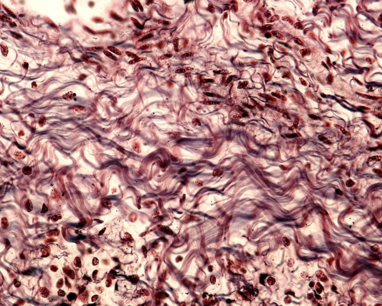
ECM is the network of molecules—including collagen, enzymes and glycoproteins—that provide structural and biochemical support to surrounding cells, including cancer cells. During the early stages of cancer, these proteins and protein fragments form the tumor microenvironment and leak into circulating blood.
“Our data reinforces the idea that this release of ECM components into circulation, even at the earliest stages of malignancy, can be used to design a specific and sensitive biomarker panel to improve detection of breast cancer,” said Pirrotte. “Using a highly specific and sensitive protein signature, we devised and verified a panel of blood-based biomarkers that could identify the earliest stages of breast cancer, and with no false positives.”
To establish this protein signature, the researchers used blood samples from 20 patients with invasive ductal carcinoma (IDC) of the breast and from 20 women without cancer who had positive mammograms but benign pathology at biopsy. These results were compared to five groups of individuals diagnosed with other cancers: ovarian, lung, prostate, colon and melanoma.
Because the number of ECM molecules in blood is relatively low, researchers relied on proteomics and new sample preparation enrichment techniques, including the use of hydrogel nanoparticles, to accurately detect cancer-associated biomarkers. This technique binds proteins from ECM associated with cancer proliferation, migration, adhesion and metastasis, or the spread of cancer from one part of the body to another. Many of these proteins had never before been observed in blood samples.
In their discovery set, 56 proteins were increased in the serum samples from IDC patients, and 32 of these proteins were specific to IDC. Verification of a subset of these proteins in an independent cohort of early-stage T1a breast cancer yielded a panel of 4 proteins, ITGA2B (integrin subunit alpha IIb), FLNA (Filamin A), RAP1A (Ras-associated protein-1A), and TLN-1 (Talin-1), which classified breast cancer patients with 100% sensitivity and 85% specificity (AUC of 0.93). These markers have been previously reported in cell-ECM interaction and tumor microenvironment biology.
“Our study results show a high degree of specificity of those markers as circulating proteins in breast cancer patients,” said Khyatiben Pathak, PhD, staff scientist in TGen’s Collaborative Center for Translational Mass Spectrometry and a study author. “Our results justify further studies with larger groups to evaluate whether this biomarker panel improves the positive predictive value of mammography for breast cancer detection.


