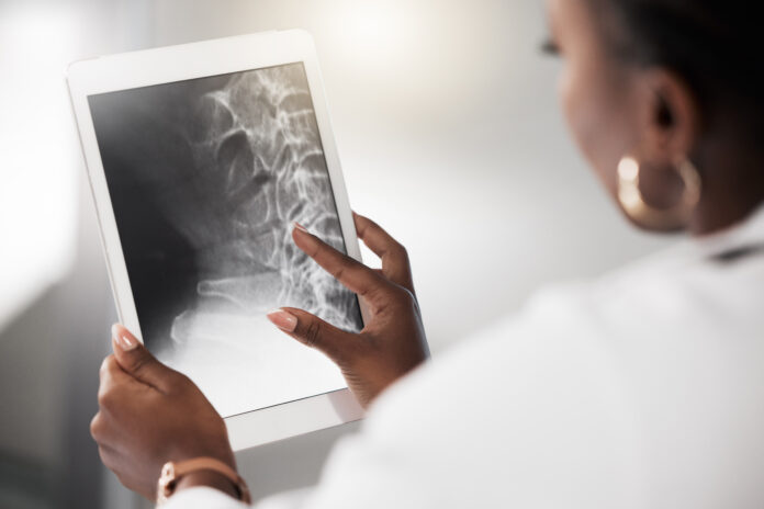
The Geneva-based Medimaps group has developed a spine segmentation (SpS) deep learning algorithm, which can enhance and improve the diagnosis and monitoring of osteoporosis.
Osteoporosis, caused by abnormally low bone density, impacts a large number of mostly older adults and is estimated to cause over 8.9 million fractures globally each year. Dual-energy X-ray absorptiometry (DXA) is currently the gold-standard technique for measuring bone density, which is the main clinical feature used to diagnose osteoporosis.
“The antero-posterior Lumbar Spine DXA scan is an important diagnostic measure, used for the assessment of osteoporosis. The quality of the scan is dependent on the accuracy of the vertebral bone mask, derived from bone edge detection and SpS,” write Guillaume Gatineau, a PhD student at the University of Lausanne and a data scientist at the Medimaps group, and colleagues in a poster presented at the European Congress of Radiology in Vienna.
“Reducing technical error requires manual validation of the default bone mask for each scan. However, this can be time-consuming in practice,” they add.
To try and assist with this problem, Gatineau and colleagues have developed a deep-learning algorithm to allow automated SpS.
To test the algorithm, the researchers selected 130 women from the OsteoLaus population cohort who had two lumbar spine DXA scans 2.5 years apart. The scans were assessed by a manufacturer default method, by the AI-based algorithm and by a clinical expert.
Following further validation tests, the team found that the AI algorithm performed better than the default analysis method for assessing bone mineral density, trabecular bone score (TBS) and surface and at a comparable level to the clinical expert for these measures.
“The volume of patients requiring scanning is steadily increasing in the radiology practice, therefore computerized tools such as the spine segmentation algorithm embedded into the TBS Osteo software represent a way forward to optimize the workflow while improving clinical patient management,” said Giuseppe Guglielmi, a professor at Foggia University School of Medicine and Dimiccoli Teaching Hospital, Barletta, Italy, in a press statement.
“This best-in class software has proven to be a precious aid in our clinical routine to detect more cases of osteoporosis, and to improve patients bone health prognostic reliably.”
The imaging software TBS Osteo, developed by Medimaps, has already received regulatory approval in the U.S. and some European countries.


