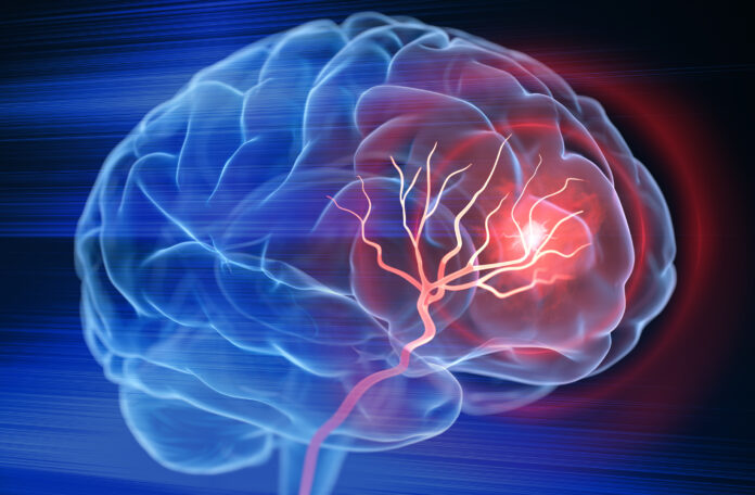
Researchers at the City University of Hong Kong (CityU) have found in a mouse study that the application of low-dose ionizing radiation (LDIR) such as X-ray radiation can reduce lesion size and reverse motor deficits in traumatic brain injury (TBI) and ischemic stroke. The method, reported in the journal Brain, Behavior, and Immunity, could provide a significant improve in the treatment of these patients as nearly half currently experience lifelong motor impairments and disability.
“Usually, secondary brain damage worsens over time after primary injuries in TBI (mechanical insults such as a car accident or falls by older adults) and strokes (when blood flow to the brain is blocked), owing to the unfavorable and hostile neuroinflammatory environment in the brain,” explained Eddie Ma Chi-him, PhD, a professor in the department of Neuroscience at CityU, who led the research. “But there is still no effective treatment for repairing the central nervous system after brain injury.”
Ma Chi-him’s team at CityU built their study based on the history of low-dose X-ray irradiation which has been shown to enhance adaptive responses that include extending life expectancy, improving immune response, improved wound healing, cell growth stimulation, and neuroprotection in animal models of neurodegenerative diseases. They theorized that given these known attributes that LDIR could potentially also have beneficial effects for TBI and stroke therapy, by mitigating damage and promoting wound healing.
The results of the study showed that not only did low-dose ionizing radiation completely reverse motor deficits and restored brain activity in TBI and stroke mouse models, but that these effects were still observed when the treatment was provided eight hours after the injury. This second finding is especially significant for translating this treatment method to the clinical as it is likely many hour may pass for patients before they could be treated.
For this research, the mice were treated for whole-body X-ray irradiation after researchers induced a brain injury or ischemic stroke, who the control mice received no irradiation. Seven days post brain injury the treated mice showed lesion size reduction of 48%, MRI showed that the treatment significantly reduced the infarct volume of the stroke mice by 43% to 51% during the first week after the stroke was induced. Past clinical observations show that stroke patients with a lower infarct volume have improved outcomes.
The treated mice also exhibited significant improvements in motor function post-treatment in both the TBI and ischemic stroke mouse populations as measured by narrow beam walking and grip strength. These measured improves showed that the LDIR mice took less time to transverse the beam and had fewer slips, evidence of motor coordination and balance.
In addition to the observed physical and function improvements, the investigators conducted transcriptomic analysis of the mice and found that the genes upregulated in the LDIR mice were enriched in pathways related to inflammatory and immune responses involving microglia. Perhaps the most important finding was that LDIR promoted axonal projections—or brain rewiring—in the motor cortex and recovered brain activity as measured by EEG months after stroke.
“Our findings indicate that LDIR is a promising therapeutic strategy for TBI and stroke patients,” Ma Chi-him concluded. “X-ray irradiation equipment for medical use is commonly available in all major hospitals. We believe this strategy could be used to address unmet medical needs in accelerating motor function restoration within a limited therapeutic window after severe brain injury, like TBI and stroke, warranting further clinical studies for a potential treatment strategy for patients.”


